patterdaleterriers.co.uk is a participant in the Amazon Services LLC Associates Program and other affiliate advertising programs designed to provide a means for us to earn fees by linking to Amazon.co.uk and affiliated sites. Affiliate links may be used on this page and in patterdaleterriers.co.uk articles, but they do not impact on the price that you pay and they do help me to get this information to you for free. Read my privacy policy for more information regarding affiliates.
Finding a lump or bump on your dog’s body can be worrying and all sorts of things can fly through your mind. Is it cancerous? How long has that been there? Is my dog in pain? Thankfully, most Histiocytomas are harmless, and your dog will recover without any side effects. Today’s blog is all about the histiocytoma in dogs.
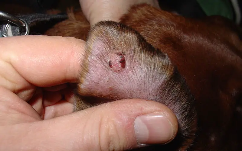
What Is a Histiocytoma?
They are small growths that appear seemingly out of nowhere usually on your dog’s head, neck, ears, or legs. They are hairless and red in colour, normally no bigger than 2.5cm in size. These growths are common patterdale terrier health problems and typically seen on younger dogs aged 3 years and under.
Dogs have cells in their body called Langerhans cells. They form a part of the immune system that fights against foreign invaders such as bacteria. Since Patterdales are natural explorers, it is no wonder they are likely to get them. Males seem to be a higher risk than females.
Histiocytomas are benign, which means they are not cancerous. They are the bodies response to a potential threat and usually resolves themselves after a month or two. However, it is better to get a vet’s opinion to rule out other illnesses.
Diagnosis of Hystiocytoma in Dogs
If your vet is confident that the lump is a histiocytoma, they will most likely suggest leaving the lump for two to three months to see if it begins to reduce in size on its own. If the lump is larger or in an awkward location, there are other options.
To be sure that the lump is a non-cancerous one, your vet will need to look at in under a microscope. That means getting hold of some cells. To do that, they may suggest a fine needle aspirate. This is a non-invasive technique used if the lump is small and in an easily accessible spot. A quick injection of local anaesthetic is given, then a needle will be used to take a sample from inside the growth.
If your dog’s growth is large or in a delicate spot such as the armpit, your vet may recommend surgical biopsy and removal. This would involve a short time under general anaesthetic so the vet can cut away all of the growth and send it away for testing.
Treatment
The most common option for treatment for hystiocytoma in dogs is to let it heal on its own. Since it is a natural reaction from the immune system, lumps like these are not harmful and tend not to cause any pain. Your vet may recommend alternative treatment if the lump has not changed within 3 months.
If your dog’s lump is a large one or is in a spot that is causing irritation, he will need to have surgery to remove it. This is usually done at the same time as a biopsy is taken, to avoid having to put the dog through multiple surgeries.
Care of your dogs Hystiocytoma
The most important thing is to keep an eye on it. If the lump is small and your vet has recommended leaving it, you will need to ensure that your dog does not lick, scratch or bite the lump as this can cause further irritation and infection.
For surgical removal, it is vital to keep your dog from licking at his wound or attempting to pull out the stitches. Gently check the temperature of the skin around the wound. Warm or hot skin can mean there is an infection. It is quite likely that your dog will have to wear the cone of shame will his skin heals!
If this blog on the histiocytoma in dogs was helpful to you, you might also like to read about maintaining Patterdale Terrier health or dog dew claw injury.